Monday, Aug. 29, 2016 | 2 a.m.
Ultrasound imaging has many purposes in medicine, and its uses continue to evolve and improve. Although many of us may associate ultrasound only with a sonogram received during pregnancy, it’s also a commonly used method to diagnose conditions throughout the entire body.
Recently, endoscopic ultrasound has started being used to diagnose diseases that have been historically difficult to detect, such as pancreatic and lung cancer. 'Endoscopic ultrasound provides very detailed images in a less invasive way,” said Tarek Ammar, MD, gastroenterology specialist at Southern Hills Hospital and Medical Center. Ultrasounds continue to be a highly valuable diagnostic tool for physicians, though their capabilities may be lesser known to most people.
How do ultrasound images work?
Ultrasounds use high-frequency sound waves to create images based on where those waves echo within the space. “The sound waves are generated through a transducer and sent to the organ or area you want an image of. The image is captured as the sound waves bounce back against the tissue,” Ammar said.
The sound waves used for ultrasounds are too high a frequency for humans to hear, and depending on the area that’s being imaged, different sound frequencies can be used. The frequencies used are usually between one and 18 megahertz. Higher-frequency waves are used to capture images with smaller, surface details, whereas lower-frequency waves are better at capturing images that involve multiple layers of tissue.
As the sound waves are pulsed into the area, the ultrasound scanner measures the echoes to create the image. The echoes are evaluated by how long they took to be received, which generally establishes the depth and location of the tissue, and how strong the echo was, which helps to establish the mass and size of the tissue.
While ultrasounds are good for creating images of soft tissue, they’re not great for capturing images of bones and structures behind bones. The physique of the patient also can affect the images produced by ultrasound. Obese patients are not good candidates for some types of traditional ultrasound because the sound waves may not be able to penetrate deeply enough into the body.
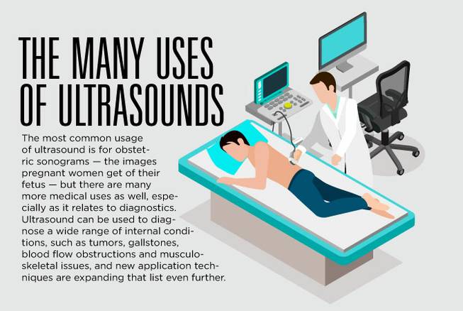
The most common usage of ultrasound is for obstetric sonograms — the images pregnant women get of their fetus — but there are many more medical uses as well, especially as it relates to diagnostics. Ultrasound can be used to diagnose a wide range of internal conditions, such as tumors, gallstones, blood flow obstructions and musculoskeletal issues, and new application techniques are expanding that list even further.
Benefits of ultrasound imaging
Among the benefits of ultrasound images is that they do not emit potentially harmful radiation, like other types of medical imaging, such as X-rays. “It is a very safe and precise imaging modality. That’s the reason why traditional ultrasound is the preferred method used during pregnancy to image the fetus without causing any harm,” Ammar said.
Other benefits to ultrasound include the ability to produce images in real time — allowing physicians to move around and explore within the area — and that the ultrasound devices themselves are portable, which allows images to be taken at a patient’s bedside.
Ultrasounds from the inside: Endobronchial and endoscopic ultrasound
Most ultrasound images are taken from outside the body, using a transducer and lubricating gel, to produce an image of what’s inside the body. Newer methods of ultrasound diagnostics, however, are taking ultrasound images from inside the body using an endoscope.
An endoscope is a flexible cable with a camera on the end, which can be inserted from the esophagus into the digestive track to look for any abnormalities. Endoscopies are nonsurgical and minimally invasive, though anesthesia is used to sedate the patient during the procedure.
An endoscopic ultrasound (EUS) is similar to a traditional endoscopy, except that it uses an ultrasound to create the images rather than a camera. Using the ultrasound produces better-quality images and can detect abnormalities that a camera may not be able to find. “The endoscopic ultrasound provides the physician more information than other imaging options,” said Ammar, who performs EUS procedures at Southern Hills Hospital and Medical Center.
EUS is used to diagnose the staging of some gastrointestinal cancers, to detect cysts and masses, and to find other abnormalities, such as lesions.
EUS also is used to provide treatment to patients. Using a fine needle at the end of the endoscope, it can draw tissue samples, insert medications, mark tumors for radiation therapy and drain cysts. The needle is guided by the ultrasound image, which is highly precise, to ensure that the correct areas are being targeted.
Similar to the EUS, endobronchial ultrasound (EBUS) is a procedure used to detect different conditions within the lungs, such as lung cancer, infections and diseases that cause enlarged lymph nodes and masses. Prior to the use of EBUS, CT scans were the most common way to collect images of the lungs, but those images tend not to be as precise or conclusive as the ones EBUS produces.
